
 |
MORPHOLOGY AND ANATOMY OF THE CALICIALES | ||||
| Thallus | Phycobionts | Apothecia | Asci | Spores | |
There are three main types of thallus in the species examined. The shape of the thallus within a single species frequently shows a wide variation.
(1) Endosubstratal thallus. The algae and hyphae are totally immersed in the substrate. When immersed in lignum, the thallus is called endoxylic.
(2) Sorediose/granular thallus. The thallus is rather thin and consists of small, ecorticate or pseudocorticate granules consisting of algae surrounded by hyphae.
(3) Verrucose/squamulose thallus. The thallus is rather thick, and the algae and hyphae are covered by a thin cortex.
Apothecia are frequent in the majority of the species with the exception of Thelomma ocellatum. Only two fertile specimens of T. ocellatum have been collected in Norway. The apothecia are stalked (in Calicium, Chaenotheca, Sclerophora, and part of the species in Microcalicium); or sessile or immersed (in Cyphelium, Thelomma, and Microcalicium disseminatum). The stalked apothecia consist of a stalk (0.33.5 mm tall) and a capitulum. There is frequently a wide variation in a single species with respect to stalk length. Some species, e.g. Calicium viride, have stout apothecia, others like Chaenotheca gracilenta have slender apothecia. The hyphae of the stalk are dark brown and irregularly arranged in Calicium and Microcalicium; and medium or pale brown and periclinally arranged in Chaenotheca and Sclerophora. The tissue containing the hymenium in an apothecium forms the excipulum. It is usually well developed, but poorly developed in some Chaenotheca and Sclerophora species. In some Sclerophora species the excipulum forms a collar. The excipulum consists of globular cells in Calicium, Cyphelium, Microcalicium and Thelomma, and elongated cells in Chaenotheca and Sclerophora. The mazaedium consists of a spore mass formed by the hymenium. The spores are released from the asci as a dry, loose powdery mass on the hymenium. This is caused by the prototunicate asci. The result is a passive dispersal of the spores, which is characteristic for all species in the Caliciales, except species in the Microcaliciaceae. The colour of the mazaedium depends on the colour of the spores. Species in Calicium, Cyphelium, and Thelomma have a black mazaedium, species in Microcalicium have a greenish black mazaedium, and species in Chaenotheca and Sclerophora have a brown mazaedium. Sometimes the mazaedium is stabilized by sclerotized hyphae (in Microcalicium). The pruina may either form a ring on the excipulum margin or it may cover the lower side of the excipulum, or even the whole stalk. The colour of the pruina may be white, brown, reddish brown or yellow (pulvinic acid derivatives). The pruina may be K+ or K-.
The asci are prototunicate and dissolve before the spores reach maturity. The spores continue maturing in the mazaedium. According to Tibell (1984), the asci lack apical structures, with the exception of species in the Microcaliciaceae. The asci may be cylindrical, clavate, or of irregular shape. They are formed singly, or, in some Chaenotheca species and in Microcalicium, in chains (catenulate). Croziers are often present in the formation of asci, but absent in Chaenotheca gracilenta and in Microcalicium. The asci contain eight spores.
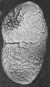
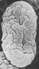
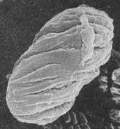
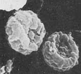
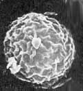
In Calicium, Cyphelium, and Thelomma, the spores are broadly ellipsoid, 8-28 [ m long, one-septate often with a constricted septum, and dark brown. In Microcalicium the spores are narrowly ellipsoid, 5-15 [ m long, 1-3-7-septate and green. In Chaenotheca and Sclerophora the spores are non-septate and pale brown; globose or ellipsoid, 5-9 [ m long in the former, and globose in the latter. In Sclerophora the spore surface is reticulate, in Chaenotheca cracked and in Microcalicium there are spirally arranged ridges. The spore surface in Calicium and Cyphelium may be more or less cracked, spirally arranged, or almost smooth. SEM photographs are shown below.
 |
 |
 |
| Calicium abietinum | Calicium glaucellum | Calicium salicinum |
 |
 |
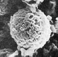 |
| Chaenotheca gracilenta | Sclerophora coniophaea | Sclerophora nivea |